|
---------------------------- 2024 Platform Presentation Winner --------------------------
Endometrial Gastric (Gastrointestinal)-Type Mucinous Adenocarcinoma (EGA): A
Clinicopathologic & Molecular Analysis of 17 Cases
Harsimar Kaur, Lawrence Hsu Lin, David L. Kolin,
Andre Pinto, Carlos Parra-Herran, Koen Van de Vijver, Glenn McCluggage, Marisa Rose Nucci
Background: EGA was introduced as a distinct entity in the latest WHO classification of female genital tumors but is rare with a
limited number of cases reported. Herein, we report the clinicopathologic, immunohistochemical and, in a
subset, molecular profiles of the largest series of EGA to date.
Design: Cases that met established criteria for EGA (gastric/gastrointestinal differentiation with supportive [at least focal
positive for gastric and/or intestinal markers] immunohistochemistry (IHC)) were confirmed from review of
available materials. Clinical data was retrieved, and histology, IHC, and available molecular results (n=7)
reviewed.
Results: The cohort consisted of 17 patients (median age 67 years; range 42 to 78 years; 13/17 postmenopausal). None
had known Peutz Jeghers syndrome (PJS). In all, bulk of the tumor was centered in the corpus (4 polypoid, 3
endophytic, 3 ill defined). Size ranged from 1.5-8.2 (mean 4.8) cm. FIGO stage ranged from IA to IVB (Table 1).
13/17 had >50% myometrial invasion, 12/17 had LVI (9/12 substantial) and 7/17 had lymph node involvement (1
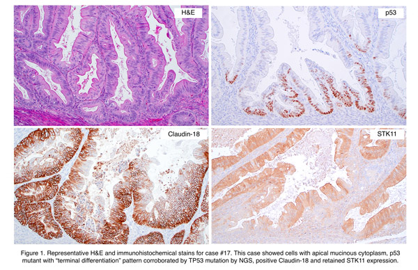
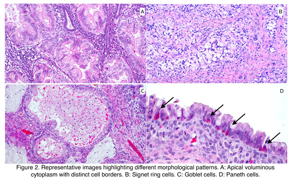
with ITC). All showed gastric/gastrointestinal differentiation: granular/foamy apical cytoplasm with distinct cell
borders (9), goblet (4), signet ring (3), Paneth (1) cells. 5 had <5% solid growth (FIGO grade 1 architecture; 8
grade 2 and 4 grade 3). Tumors were positive for MUC6 (7/13), CK20 (8/12), CDX2 (15/16) and Claudin 18 (4/7).
10/17 were p53 mutant (2 with “terminal differentiation”) and 2/14 showed MLH1 and PMS2 loss with MLH1
gene promoter methylation. STK11 was retained in 7/8 and equivocal in 1. HER2 was 3+ positive with ERBB2
amplification (1), 2+ (5) and 0 (1).
Next generation sequencing showed pathogenic variants in TP53 (7/7), KRAS (3/7), BRAF (1/7), and SMAD4
(1/7). Outcome data (13 patients; 2-48 months) showed: 1 patient dead of disease 14 mo after diagnosis; 8 alive
with disease including 1 with multi-site metastasis (14 mo) and 1 with lung metastasis (48 mo); and 4 patients
alive with no evidence of disease (9-47 mo).
Results Table:
| Case # |
Age |
Presenting symptom |
Location of tumor |
FIGO Stage |
% myometrial invasion |
LVSI |
Relevant POSITIVE IHC results |
Relevant NEGATIVE IHC results |
| 1 |
50 |
Intermenstrual bleeding |
LUS |
IA |
None |
No |
PAX8, MUC6, ER (weak), CDX2 |
CK20, PR, p53 wild-type, MMR (intact) |
| 2 |
67 |
Postmenopausal bleeding, pelvic pain |
Fundus |
IB |
More than 50% |
Yes, substantial |
CK20, CDX2, loss of MLH1 and PMS2 |
PAX8, ER, PR, p53 wild-type |
| 3 |
78 |
Urinary symptoms |
Fundus and body |
IB |
More than 50% |
Yes, substantial |
PAX8, CDX2, p53 mutant |
MUC6, ER, MMR (intact) |
| 4 |
87 |
NA |
Endometrium |
IB |
More than 50% |
No |
PAX8, MUC6, Claudin 18, CDX2, ER (focal), PR (focal), p53 mutant (terminal differentiation), HER2 (2+) |
|
| 5 |
44 |
Fibroid |
LUS |
IB |
More than 50% |
Yes, substantial |
PAX8, CDX2, ER (rare), p16 (diffuse), p53 mutant |
PR |
| 6 |
71 |
Postmenopausal bleeding |
Fundus |
IB |
More than 50% |
No |
PAX8, GATA3, CDX2, HNF1beta |
ER, PR, CK20, MUC6, p53 wild-type, MMR (intact) |
| 7 |
66 |
NA |
Endometrium and invasion of cervical stroma |
II |
More than 50% |
Yes |
PAX8, p16 (diffuse), Napsin A, MUC6, CDX2, Claudin 18, mucicarmine, p53 mutant (null-pattern), HER2 (2+) |
ER, PR, CK20, MMR (intact) |
| 8 |
64 |
Bilateral complex masses |
Endometrium, bilateral ovaries, and fallopian tubes |
IIIA |
None |
No |
CDX2, CK20, p53 mutant |
PAX8, ER, PR, MMR (intact) |
| 9 |
42 |
NA |
Endometrium, right adnexa |
IIIA |
None |
No |
CDX2, MUC6, Subclonal loss of ARID1A, p53 mutant |
CK20, PTEN (retained) |
| 10 |
73 |
Postmenopausal bleeding |
Fundus and body |
IIIB |
More than 50% |
Yes, focal |
CK20, CDX2, MUC6, loss of MLH1 and PMS2 |
PAX8, ER, PR, p53 wild-type |
| 11 |
71 |
Postmenopausal bleeding |
Fundus and body |
IIIC1 |
More than 50% |
Yes, substantial |
MUC6, Claudin 18, CK20, CDX2, p53 mutant |
ER, PR, HER2 (0), MMR (intact) |
| 12 |
69 |
Postmenopausal bleeding, abdominal pain |
Body |
IIIC1 |
More than 50% |
Yes, substantial |
P53 mutant, AMACR, Napsin A |
ER, PR, MMR (intact) |
| 13 |
75 |
Postmenopausal bleeding |
Endometrium, bilateral ovaries, and right fallopian tubes |
IIIC1 |
More than 50% |
Yes, substantial |
PAX8, CK20, mucicarmine, CDX2, p53 mutant (overexpressed), HER2 (2+) |
ER, PR, MUC6, Claudin 18, MMR (intact) |
| 14 |
62 |
Postmenopausal bleeding |
Body |
IIIC2 |
More than 50% |
Yes, substantial |
PAX8, ER (rare) |
MUC6, CDX2, p53 wild-type, MMR (intact) |
| 15 |
75 |
Postmenopausal bleeding |
Body (multifocal) |
IIIC2 |
More than 50% |
Yes |
CDX2, CK20, HER2 (2+) |
PAX8, ER, PR, MUC6, Claudin 18, p53 wild-type, MMR (intact) |
| 16 |
67 |
NA |
Endometrium, cervical stroma, and right adnexa |
IIIC2 |
More than 50% |
Yes, substantial |
CK20, CDX2, p16 (diffuse), HER2 (3+), PD-L1 >1 |
PAX8, ER, PR, MUC6, Claudin18, HPV ISH, p53 wild-type, MMR (intact) |
| 17 |
48 |
Pelvic pain |
LUS, bilateral ovaries and fallopian tubes, peritoneum |
IVB |
Less than 50% |
Yes, substantial |
PAX8, MUC6, Claudin18, CK20, CDX2, p53 mutant (terminal differentiation), HER2 (2+) |
ER, PR, MMR (intact) |
Conclusions: Like their cervical counterpart, primary EGA have a distinctive morphologic appearance, harbor frequent TP53
mutations and can be associated with adverse outcome despite well differentiated glandular morphology and/or
low stage disease. In contrast, they do not appear to be associated with PJS or STK11 alterations, although
number of cases studied is small.
|